
Ficheiro:Nanotubes.png

Dimensões desta antevisão: 782 × 600 píxeis. Outras resoluções: 313 × 240 píxeis | 626 × 480 píxeis | 1 002 × 768 píxeis | 1 280 × 982 píxeis | 2 128 × 1 632 píxeis.
Imagem numa resolução maior (2 128 × 1 632 píxeis, tamanho: 2,29 MB, tipo MIME: image/png)
|
|
Esta imagem provém do Wikimedia Commons, um acervo de conteúdo livre da Wikimedia Foundation que pode ser utilizado por outros projetos.
|
Descrição do ficheiro
| Descrição |
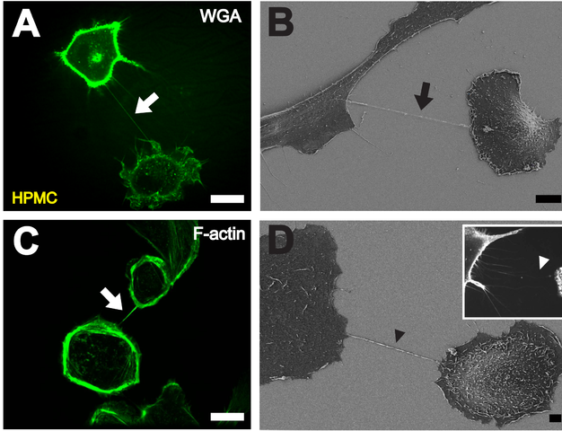
English: A. High resolution 3D live-cell fluorescence image of a NT (white arrow) connecting two primary mesothelial cells one hour after plating on a collagen I coated glass cover slide. To facilitate detection, cell membranes were stained with WGA Alexa Fluor® 488. Scale bar: 20 µm. B Depiction of a NT (black arrow) between two cells with scanning electron microscopy one hour after cell plating. Scale bar: 10 µm. C F-actin staining by fluorescently labeled phalloidin showing actin being present in NTs between individual HPMCs (white arrow). Scale bar: 20 µm. D Scanning electron microscope picture of a substrate-associated filopodia-like extension as potential NT precursor (black arrowhead). The insert shows a fluorescence microscopic image of substrate associated filopodia-like protrusions approaching a neighboring cell (white arrowhead). Scale bar: 2 µm. |
| Data | |
| Origem | PLoS One |
| Autor | Ranzinger J, Rustom A, Abel M, Leyh J, Kihm L, et al. |
Licenciamento
|
A utilização deste ficheiro é regulada nos termos da licença Creative Commons - Atribuição 2.5 Genérica.
|
This file was published in a Public Library of Science journal. Their website states that the content of all PLOS journals is published under the Creative Commons Attribution 4.0 license (or its previous version depending on the publication date), unless indicated otherwise.
|
Histórico do ficheiro
Clique uma data e hora para ver o ficheiro tal como ele se encontrava nessa altura.
| Data e hora | Miniatura | Dimensões | Utilizador | Comentário | |
|---|---|---|---|---|---|
| atual | 11h44min de 28 de dezembro de 2011 | | 2 128 × 1 632 (2,29 MB) | Gustavocarra | {{Information |Description ={{en|1='''A'''. High resolution 3D live-cell fluorescence image of a NT (white arrow) connecting two primary mesothelial cells one hour after plating on a collagen I coated glass cover slide. To facilitate detection, cell me |
Utilização local do ficheiro
A seguinte página usa este ficheiro:
Utilização global do ficheiro
As seguintes wikis usam este ficheiro:
- ar.wikipedia.org
- en.wikipedia.org
- es.wikipedia.org
- gl.wikipedia.org
- ja.wikipedia.org