
Ficheiro:Fundus of patient with retinitis pigmentosa, mid stage.jpg
Dimensões desta antevisão: 699 × 599 píxeis. Outras resoluções: 280 × 240 píxeis | 560 × 480 píxeis | 871 × 747 píxeis.
Imagem numa resolução maior (871 × 747 píxeis, tamanho: 107 kB, tipo MIME: image/jpeg)
|
|
Esta imagem provém do Wikimedia Commons, um acervo de conteúdo livre da Wikimedia Foundation que pode ser utilizado por outros projetos.
|
| Descrição |
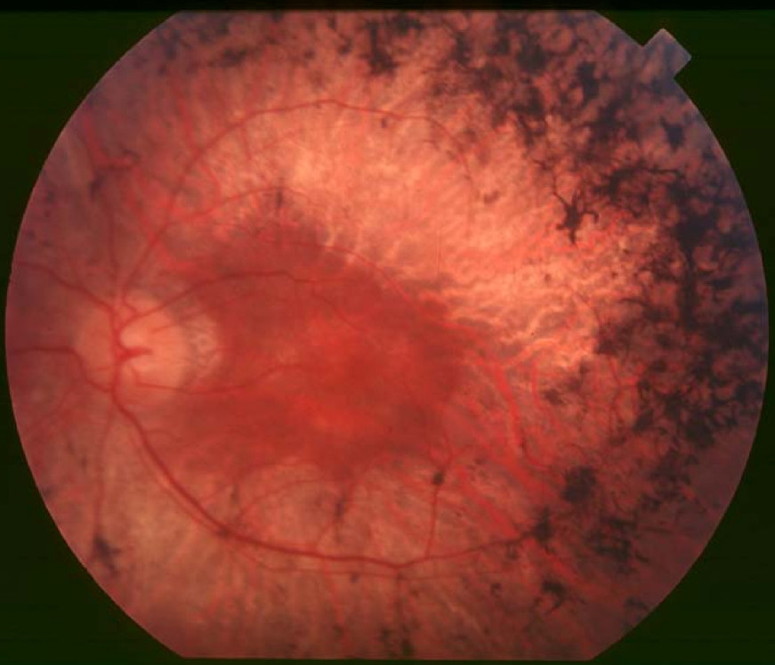
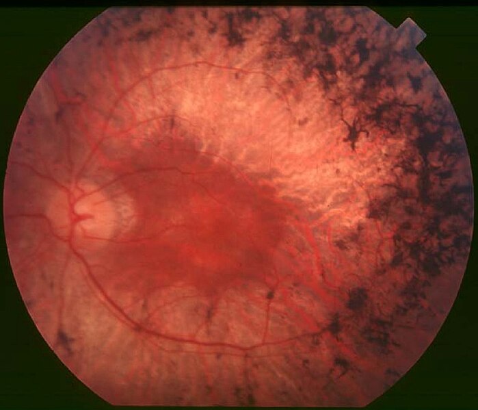
English: Figure 2. Fundus of patient with retinitis pigmentosa, mid stage (Bone spicule-shaped pigment deposits are present in the mid periphery along with retinal atrophy, while the macula is preserved although with a peripheral ring of depigmentation. Retinal vessels are attenuated.) Hamel Orphanet Journal of Rare Diseases 2006 1:40 doi:10.1186/1750-1172-1-40 |
| Data | |
| Origem | Retinitis pigmentosa by Christian Hamel |
| Autor | Christian Hamel |
| Permissão (Reutilizar este ficheiro) |
© 2006 Hamel; licensee BioMed Central Ltd. This is an Open Access article distributed under the terms of the Creative Commons Attribution License (https://creativecommons.org/licenses/by/2.0), which permits unrestricted use, distribution, and reproduction in any medium, provided the original work is properly cited. |
A utilização deste ficheiro é regulada nos termos da licença Creative Commons - Atribuição 2.0 Genérica.
- Pode:
- partilhar – copiar, distribuir e transmitir a obra
- recombinar – criar obras derivadas
- De acordo com as seguintes condições:
- atribuição – Tem de fazer a devida atribuição da autoria, fornecer uma hiperligação para a licença e indicar se foram feitas alterações. Pode fazê-lo de qualquer forma razoável, mas não de forma a sugerir que o licenciador o apoia ou subscreve o seu uso da obra.
Histórico do ficheiro
Clique uma data e hora para ver o ficheiro tal como ele se encontrava nessa altura.
| Data e hora | Miniatura | Dimensões | Utilizador | Comentário | |
|---|---|---|---|---|---|
| atual | 10h17min de 2 de dezembro de 2017 | | 871 × 747 (107 kB) | Doc James | Cropped 27 % horizontally and 7 % vertically using CropTool with precise mode. |
| 13h52min de 22 de setembro de 2009 |  | 1 200 × 799 (126 kB) | CopperKettle | {{Information |Description={{en|1=Figure 2. Fundus of patient with retinitis pigmentosa, mid stage (Bone spicule-shaped pigment deposits are present in the mid periphery along with retinal atrophy, while the macula is preserved although with a peripheral |
Utilização local do ficheiro
As seguintes 2 páginas usam este ficheiro:
Utilização global do ficheiro
As seguintes wikis usam este ficheiro:
- ar.wikipedia.org
- bs.wikipedia.org
- ca.wikipedia.org
- da.wikipedia.org
- en.wikipedia.org
- en.wikiversity.org
- es.wikipedia.org
- eu.wikipedia.org
- fa.wikipedia.org
- fi.wikipedia.org
- fr.wikipedia.org
- he.wikipedia.org
- hy.wikipedia.org
- it.wikipedia.org
- ko.wikipedia.org
- la.wikipedia.org
- or.wikipedia.org
- outreach.wikimedia.org
- pl.wikipedia.org
- ru.wikipedia.org
- sl.wikipedia.org
- sr.wikipedia.org
- sv.wikipedia.org
- th.wikipedia.org
- tr.wikipedia.org
- tt.wikipedia.org
- uk.wikipedia.org
- vi.wikipedia.org
- www.wikidata.org