
Ficheiro:Echinococcus Life Cycle.svg

Dimensões desta antevisão em PNG do ficheiro SVG: 629 × 600 píxeis Outras resoluções: 252 × 240 píxeis | 504 × 480 píxeis | 806 × 768 píxeis | 1 074 × 1 024 píxeis | 2 149 × 2 048 píxeis | 1 280 × 1 220 píxeis.
Imagem numa resolução maior (ficheiro SVG, de 1 280 × 1 220 píxeis, tamanho: 643 kB)
|
|
Esta imagem provém do Wikimedia Commons, um acervo de conteúdo livre da Wikimedia Foundation que pode ser utilizado por outros projetos.
|
Descrição do ficheiro
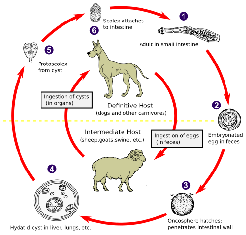
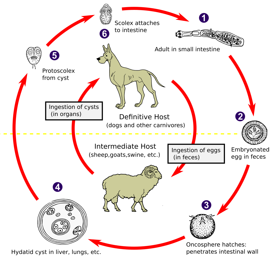
| Descrição | The adult Echinococcus granulosus (3 to 6 mm long) [1] resides in the small bowel of the definitive hosts (dogs or other carnivores). Gravid proglottids release eggs [2] that are passed in the feces. After ingestion by a suitable intermediate host (under natural conditions: sheep, goat, swine, cattle, horses, camel), the egg hatches in the small bowel and releases an oncosphere [3] that penetrates the intestinal wall and migrates through the circulatory system into various organs, especially the liver and lungs. In these organs, the oncosphere develops into a cyst [4] that enlarges gradually, producing protoscolices and daughter cysts that fill the cyst interior. The definitive host becomes infected by ingesting the cyst-containing organs of the infected intermediate host. After ingestion, the protoscolices [5] evaginate, attach to the intestinal mucosa [6] and develop into adult stages [1] in 32 to 80 days. The same life cycle occurs with E. multilocularis (1.2 to 3.7 mm), with the following differences: the definitive hosts are foxes, and to a lesser extent dogs, cats, coyotes and wolves; the intermediate host are small rodents; and larval growth (in the liver) remains indefinitely in the proliferative stage, resulting in invasion of the surrounding tissues. With E. vogeli (up to 5.6 mm long), the definitive hosts are bush dogs and dogs; the intermediate hosts are rodents; and the larval stage (in the liver, lungs and other organs) develops both externally and internally, resulting in multiple vesicles. E. oligarthrus (up to 2.9 mm long) has a life cycle that involves wild felids as definitive hosts and rodents as intermediate hosts. Humans become infected by ingesting eggs , with resulting release of oncospheres in the intestine and the development of cysts in various organs. Image adapted from original available at the United States Centres for Disease Control Parasitology Identification Laboratory ([1]). |
| Data | |
| Origem |
Este ficheiro foi derivado de: Echinococcus Life Cycle.png: |
| Autor |
CDC Vetor: 🎱 |
| SVG desenvolvimento | Este diagrama inválido foi criado com o Other tools |
Licenciamento
Esta imagem é um trabalho dos Centers for Disease Control and Prevention, parte do Departamento de Saúde e Serviços Humanos dos Estados Unidos da América, tirada ou feita durante o curso de uma tarefa oficial de um funcionário. Como trabalho do Governo Federal dos Estados Unidos da América, a imagem está no domínio público.
|
Registo de carregamento original
This image is a derivative work of the following images:
- Echinococcus Life Cycle.png licensed with PD-USGov-HHS-CDC
- 2007-01-24T10:54:56Z Pngbot 600x571 (45555 Bytes) optimized with optipng
- 2005-04-26T01:48:50Z FirstPrinciples~commonswiki 600x571 (55999 Bytes) Smaller & clearer
- 2005-04-26T01:36:23Z FirstPrinciples~commonswiki 800x761 (80990 Bytes)
Carregada com derivativeFX
Histórico do ficheiro
Clique uma data e hora para ver o ficheiro tal como ele se encontrava nessa altura.
| Data e hora | Miniatura | Dimensões | Utilizador | Comentário | |
|---|---|---|---|---|---|
| atual | 01h31min de 1 de fevereiro de 2021 | | 1 280 × 1 220 (643 kB) | Pixelsquid | Resized. |
| 20h44min de 31 de janeiro de 2021 |  | 320 × 305 (460 kB) | Pixelsquid | == {{int:filedesc}} == {{Information |Description=The adult Echinococcus granulosus (3 to 6 mm long) [1] resides in the small bowel of the definitive hosts (dogs or other carnivores). Gravid proglottids release eggs [2] that are passed in the feces. After ingestion by a suitable intermediate host (under natural conditions: sheep, goat, swine, cattle, horses, camel), the egg hatches in the small bowel and releases an oncosphere [3] that penetrates the intestinal wall and migrates through the... |
Utilização local do ficheiro
A seguinte página usa este ficheiro:
Utilização global do ficheiro
As seguintes wikis usam este ficheiro:
- ar.wikipedia.org
- arz.wikipedia.org
- be.wikipedia.org
- bs.wikipedia.org
- ca.wikipedia.org
- dag.wikipedia.org
- el.wikipedia.org
- en.wikipedia.org
- es.wikipedia.org
- fa.wikipedia.org
- ga.wikipedia.org
- gl.wikipedia.org
- hi.wikipedia.org
- hu.wikipedia.org
- hy.wikipedia.org
- ia.wikipedia.org
- id.wikipedia.org
- is.wikipedia.org
- it.wikipedia.org
- ja.wikipedia.org
- ko.wikipedia.org
- ky.wikipedia.org
- lt.wikipedia.org
- mk.wikipedia.org
- ml.wikipedia.org
- ms.wikipedia.org
- nl.wikipedia.org
- om.wikipedia.org
- or.wikipedia.org
- pl.wikipedia.org
- ro.wikipedia.org
- ru.wikipedia.org
- sl.wikipedia.org
- sr.wikipedia.org
- sv.wikipedia.org
- th.wikipedia.org
- tl.wikipedia.org
- tr.wikipedia.org
- uk.wikipedia.org
- uz.wikipedia.org
- vi.wikipedia.org
- www.wikidata.org
Ver mais utilizações globais deste ficheiro.